Research Theme

Reactive transport & CO2 Storage

How growing carbonate minerals fracture ultramafic rock and connect permanent carbon storage with lower-energy mineral extraction.

01

Background

Carbon mineralization stores carbon dioxide by converting it into solid carbonate minerals. Magnesium- and calcium-rich rocks, including basalt and peridotite, contain many of the elements needed for this reaction. Once formed, carbonate minerals can retain carbon for geologic time. Projects in Iceland and Washington State have already demonstrated mineral storage in basalt, while natural carbonate veins in ultramafic rocks record the same process over much longer periods.

The challenge is fluid access. Many ultramafic rocks contain abundant reactive minerals but transmit fluids slowly. Mineral growth can narrow pores and seal fractures, limiting how much rock reacts. Growth inside a confined vein can also push against the surrounding rock, create cracks, expose fresh mineral surfaces, and open new pathways for CO2-rich water. This reaction-driven cracking may sustain carbon mineralization through a self-reinforcing cycle of reaction, expansion, fracture growth, and renewed fluid access.

The same process could help extract critical minerals. Ultramafic rocks can host nickel and other metals, yet crushing and grinding them consumes substantial energy. Carbonation that fractures and weakens the rock could permanently store CO2 while preparing the ore for mining. This study tests that possibility by reacting brucite-rich ultramafic rock with carbonated, magnesium-rich water under elevated pressure and temperature, then tracking where minerals formed and how the rock deformed.

02

Questions driving this work

  • How quickly can CO2 mineralization create fractures and weaken ultramafic rock?
  • Where do new cracks begin, and how do pre-existing brucite veins guide their growth?
  • Which newly formed minerals occupy the reacted veins and fracture surfaces?
  • Can mineral growth generate enough pressure to overcome the rock's strength and fracture toughness?
  • How do temperature and magnesium availability control dissolution, precipitation, and damage?
  • Could reaction-driven cracking sustain fluid access for carbon storage while reducing the energy needed to process mineral ores?

03

Materials and methods

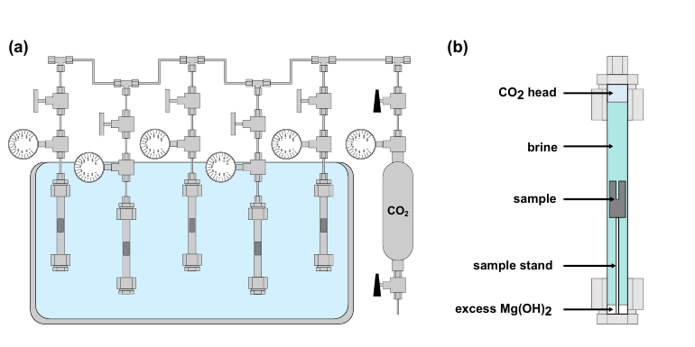
Diagram of pressurized reactor tubes in a temperature-controlled bath and a rock sample submerged in magnesium-rich brine beneath a carbon dioxide headspace.
Supporting Figure 1. Pressurized reactor tubes held the rock samples in hot, magnesium-rich brine beneath a CO2 headspace.

Rock samples and reaction conditions

The samples came from the Crawford Ultramafic Complex in Ontario, Canada, and were collected from 129.8 to 132.0 meters below the surface. The rock is serpentinized dunite: an ultramafic rock composed mainly of serpentine minerals, small remnants of olivine, brucite-rich veins, and trace magnetite. Each cylindrical mini-plug measured 9.5 millimeters across and 21 millimeters long. A narrow blind hole increased the reactive surface area and connected the fluid with internal veins and flaws.

Stainless-steel reactors held the samples at 90°C and 3.5 megapascals of CO2 pressure. The fluid contained deionized water and magnesium hydroxide, which maintained magnesium-rich conditions comparable to water that has already reacted with brucite farther from an injection well. The main time series lasted 2, 4, 11, 18, and 32 days. Two 11-day controls isolated key chemical effects: carbonated water at 90°C and magnesium-rich carbonated water at 20°C. The samples experienced fluid pressure with no externally applied confining load, creating low effective-stress conditions.
Three-dimensional imaging and deformation

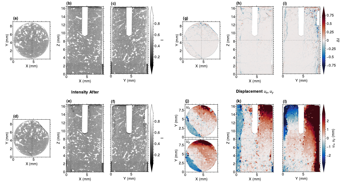
Micro-computed X-ray tomography scanned each recoverable sample before and after reaction at a voxel size of 12.7 micrometers. A voxel is a three-dimensional pixel. After aligning the scans, digital volume correlation tracked small patterns within the rock from one scan to the next. This process works like feature tracking in a video and produces a three-dimensional displacement field. The resulting maps identify where the rock expanded, contracted, separated, or fractured.

Mineral identification and fracture mechanics

Raman spectroscopy identified minerals from the way they scatter laser light. Scanning electron microscopy with energy-dispersive X-ray spectroscopy mapped elements at much smaller scales. Together, these tools connected mineral changes with the veins and fractures identified in the X-ray scans. Semi-circular bending tests measured the fracture toughness of related rock samples. A coupled thermodynamic and fracture-mechanics model then estimated the pressure generated by carbonate growth and the minimum flaw size that could propagate as a crack.

04

Results

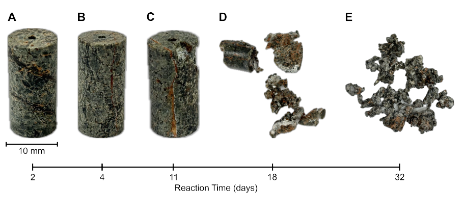
Ultramafic rock mini-plugs after 2, 4, 11, 18, and 32 days of reaction, showing progressive cracking and disaggregation.
Figure 1. Visible damage appeared by day 11. The rock broke into progressively smaller fragments after 18 and 32 days.
  • Damage developed rapidly and progressed with time. The 2- and 4-day samples showed subtle surface alteration and little organized internal deformation. Cracks and detached fragments appeared by day 11. The 18-day sample broke into pieces smaller than about 5 millimeters, and the 32-day sample disaggregated into millimeter-scale fragments with little cohesive strength.
  • Expansion concentrated along veins and flaw tips. Three-dimensional image analysis revealed outward displacement beside pre-existing brucite veins and newly formed fractures. The strongest changes followed mineral pathways that connected the brine with the rock interior.
  • The controls isolated the importance of chemistry and temperature. Carbonated water at 90°C produced little coherent deformation when the fluid lacked the magnesium hydroxide buffer. The 20°C magnesium-rich experiment produced dissolution and modest deformation, with far less cracking than the 90°C experiment.
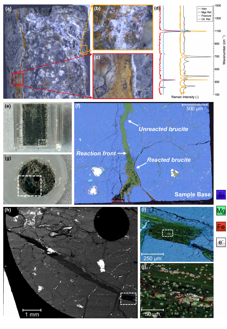
  • Magnesite formed within reacted brucite veins. Raman spectroscopy and elemental maps identified magnesium carbonate beside the veins and on newly exposed fracture surfaces. Rounded, iron-bearing magnesite grains occupied altered parts of the original vein and were surrounded by dense microfractures.
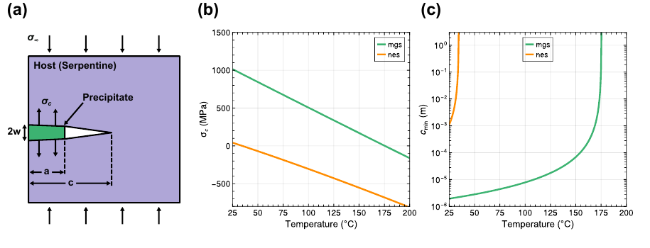
  • Mineral growth generated enough pressure to propagate small cracks. The model predicts magnesite crystallization pressures above 100 megapascals. At 90°C, this pressure can extend a pre-existing crack about 10 micrometers long, consistent with the measured fracture toughness of the rock.
  • Carbonation created a feedback between chemistry and mechanics. Mineral growth expanded confined veins, opened fractures, exposed fresh surfaces, and improved access for additional reactive fluid. This feedback can help mineralization advance through otherwise low-permeability rock.
  • The reaction also weakened the rock. Progressive fracturing and disaggregation suggest that CO2-rich fluids could mechanically prepare ultramafic ore for crushing and mineral separation while storing carbon as solid carbonate.
X-ray slices before and after reaction, intensity-difference maps, and three-dimensional displacement maps for the 11-day sample.
Figure 2. X-ray images document the rock before and after 11 days of reaction. Difference maps locate material changes, while digital volume correlation reveals outward movement beside veins and fractures.
Optical images, Raman spectra, and electron-microscope elemental maps locating reacted brucite, a carbonate reaction front, and iron-bearing magnesite near new fractures.
Figure 3. Raman spectra and elemental maps identify magnesite within altered brucite veins and beside newly formed fractures.

Connecting mineral growth with fracture formation

The mineral maps place the reaction front directly inside the damaged zones identified by X-ray imaging. Magnesite replaced part of a brucite-rich vein near the sample base and formed rounded grains with iron-rich cores and magnesium-rich rims. Fractures spread through the reacted vein and into the surrounding serpentine matrix. The close spatial match among magnesite, local expansion, and cracking supports mineral growth as the principal driver of damage.

Conceptual model of carbonate precipitation pushing open a crack, with graphs of crystallization pressure and minimum crack size versus temperature.
Supporting Figure 7. Confined magnesite growth can exert more than 100 megapascals of pressure and propagate micron-scale flaws at the experimental temperature.

A physically plausible cracking mechanism

The experiments recorded strong local expansion along reacted veins. When magnesite grows inside a confined flaw, its growth pushes against the crack walls. The calculated pressure exceeds the measured strength and fracture toughness of the serpentinized dunite. The model also indicates that shallow crustal confinement would reduce the driving stress only slightly, making this mechanism relevant to depths that engineered wells can reach.

05

Future directions

  • Apply realistic subsurface stresses. Repeat the experiments under controlled confining and differential stress to determine when burial pressure suppresses or redirects reaction-driven cracks.
  • Measure permeability during reaction. Flow-through experiments could track injectivity, fluid pathways, and the competition between fracture opening and mineral sealing as they evolve.
  • Resolve the onset of cracking. Repeated or in-situ X-ray scans could identify the first growing crystals, measure crack speed, and test whether fracture growth accelerates the chemical reaction.
  • Vary rock and fluid composition. Experiments across brucite abundance, vein geometry, salinity, pH, temperature, CO2 delivery method, and common fluid impurities would define the conditions that favor sustained cracking.
  • Strengthen the coupled model. Reactive-transport simulations can incorporate the measured crystallization pressure, fracture toughness, evolving surface area, fluid flow, and precipitation kinetics.
  • Quantify mining benefits. Larger samples could measure changes in compressive strength, crushing energy, grinding energy, and nickel recovery after controlled carbonation.
  • Test field-scale performance and storage security. Pilot injections could determine how fracture networks spread, how much CO2 mineralizes, whether pathways remain connected, and how deformation or microseismicity develops.